AIIMS Doctors Identify Two High-Risk Zones to Improve Safety in Complex Cancer Surgeries
Study of 255 cancer operations highlights critical anatomical areas where careful dissection may help limit bleeding and protect the urinary tract, pelvic nerves and major blood vessels.
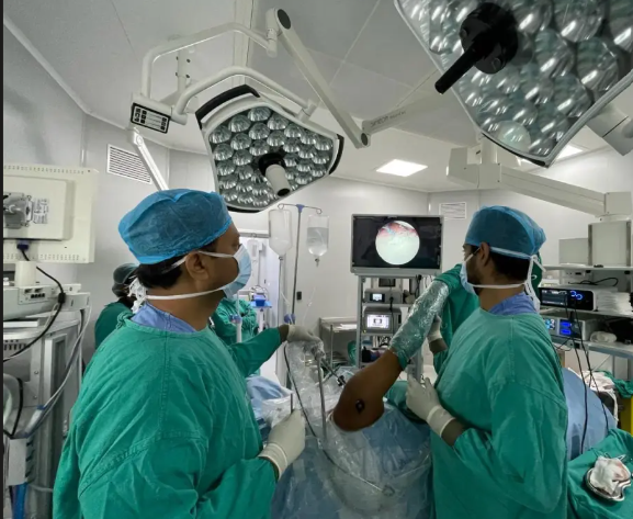
NEW DELHI, Oct 19: Doctors at the All India Institute of Medical Sciences (AIIMS), New Delhi, have identified two high-risk anatomical areas that could help surgeons reduce complications during complex cancer operations, particularly procedures involving the reproductive organs and liver.
The surgical concepts, developed by Dr M D Ray, a professor in the Department of Surgical Oncology at the Dr B R Ambedkar Institute Rotary Cancer Hospital (Dr BRA-IRCH), and his team, focus on recognising vulnerable structures before operating in areas where even minor errors can cause serious bleeding or damage to vital organs.
One of the areas, described as the “Red Alert Zone” or “Ray’s Zone”, lies below the uterine blood vessels in the pelvis. The other, called “Ray’s Hepatocaval Triangle”, provides an anatomical reference for surgeons working around the liver and major blood vessels.
The identification of these areas is intended to encourage greater precision during technically demanding procedures. Researchers say the approach may help reduce blood loss and lower the risk of injuries to the ureter, urinary bladder, pelvic nerves and major vascular structures.
The findings, reported on October 18, are based on an analysis of 255 radical or modified radical hysterectomies performed for cervical, endometrial and ovarian cancers. The team examined surgical anatomy and the causes of bleeding and injuries before modifying its approach in the later 105 operations.
Identifying the Pelvic Region Where Errors Can Have Serious Consequences
Cancer surgery involving the uterus and surrounding tissues can be technically challenging because several important structures lie close together in a confined space.
The ureter, which carries urine from the kidneys to the bladder, passes through the pelvis near blood vessels and other tissues that surgeons may need to examine or remove during an operation. The urinary bladder and nerves responsible for pelvic functions can also lie close to the surgical field.
When cancer treatment requires extensive dissection, these structures must be carefully identified and protected. Damage to a blood vessel can cause significant bleeding, while injury to the urinary tract or pelvic nerves may lead to complications that affect recovery and quality of life.
Ray’s Zone draws attention to this vulnerable region and encourages surgeons to approach it with particular care. Rather than treating the area as an ordinary part of the operative field, the concept highlights the need to establish the precise location of surrounding structures before proceeding.
The idea emerged from the team’s examination of 255 complex cancer operations. By reviewing anatomical relationships and the circumstances associated with operative bleeding or injury, the researchers sought to identify where additional precautions could improve surgical safety.
The approach was subsequently modified in 105 cases, allowing the team to apply more deliberate techniques in the identified high-risk area.
The work highlights how detailed anatomical understanding can influence surgical practice. While advanced instruments and imaging technologies can assist surgeons, safe operating also depends on recognising vulnerable structures and understanding their relationship to the tissues being treated.
Greater Emphasis on Direct Visualisation
The revised approach places importance on carefully exposing and identifying anatomical structures before cutting or separating tissues.
Surgeons may use magnification when necessary to improve visibility in confined areas. Individual blood vessels can be identified and tied off separately rather than relying on broad tissue handling that could obscure important structures.
The technique also emphasises avoiding excessive cautery near the ureter. Cautery uses heat to cut tissue or control bleeding, but unintended thermal injury can damage nearby structures even when there is no obvious direct cut.
Careful control of instruments and energy devices is therefore particularly important when several vital structures lie close together.
The researchers said meticulous dissection in Ray’s Zone could help minimise blood loss and reduce the likelihood of injuries involving the ureter, bladder and pelvic nerves.
Such precautions are relevant not only during the operation but also to the patient’s recovery. Complications involving the urinary tract or nerves may require additional treatment and can affect normal bodily functions after surgery.
Reducing avoidable injury can help support better postoperative outcomes, although the result of any individual procedure also depends on the patient’s condition, the extent of the disease and the complexity of the operation.
The concept is not a replacement for established surgical training or clinical judgment. Instead, it provides a specific anatomical warning point that may help surgeons organise their approach to a difficult part of an operation.
A Second Landmark Near the Liver
The team’s other concept, Ray’s Hepatocaval Triangle, concerns a different but equally demanding surgical region near the liver.
The area becomes important during liver mobilisation and dissection around the inferior vena cava and major hepatic veins. These vessels carry blood through the liver and are essential to normal circulation.
Operations involving this region can be challenging because major vascular structures may be close to the tissues being manipulated. Uncontrolled injury can result in severe bleeding and require immediate intervention.
Identifying a clearly defined anatomical area can provide surgeons with a reference point before they begin dissection. The aim is to encourage controlled movements and careful identification of structures rather than proceeding without a clear understanding of the local anatomy.
The concept shares the same underlying principle as Ray’s Zone: recognise the part of the body where the consequences of an error could be particularly serious, then plan the surgical approach around protecting the structures located there.
Although the two zones are situated in different parts of the body and apply to different operative situations, both emphasise the importance of anatomical precision in complex surgery.
Why Blood Loss Remains a Major Surgical Concern
Controlling bleeding is a fundamental consideration in major cancer operations. Significant blood loss can complicate the procedure, require additional interventions and increase the demands placed on the surgical and anaesthesia teams.
The risk may be greater when a tumour lies close to major blood vessels or when surgeons need to remove extensive tissue to treat the disease.
Preventive measures include careful identification of vessels, deliberate tissue separation and the use of appropriate techniques to control bleeding. Surgeons must also be prepared to respond quickly if an unexpected injury occurs.
The AIIMS team’s emphasis on individual vessel ligation and controlled dissection is consistent with the broader objective of reducing avoidable damage during difficult operations.
Protecting nearby organs is equally important. A technically successful removal of cancerous tissue must be balanced against the need to preserve urinary function, nerve function and other essential physiological processes wherever possible.
For patients, fewer complications may mean a smoother recovery and a lower likelihood of requiring additional procedures. However, the benefits of a particular technique must be assessed in the context of the patient’s diagnosis and the complexity of the surgery.
Importance for Gynaecological Cancer Treatment
Cervical, endometrial and ovarian cancers may require operations involving the uterus, surrounding tissues and nearby anatomical structures. The precise procedure depends on the type of cancer, its stage and whether it has spread to other areas.
Radical hysterectomy involves removing the uterus and additional surrounding tissues, while modified radical procedures may involve a different extent of tissue removal depending on the clinical circumstances.
Because these operations can require dissection close to blood vessels, urinary structures and nerves, a detailed understanding of pelvic anatomy is essential.
Ray’s Zone may serve as a practical reminder during these procedures that several important structures occupy a limited space. The approach encourages surgeons to identify them individually and avoid unnecessary tissue injury.
The research is particularly relevant to surgical oncology, where treatment often requires a balance between removing diseased tissue adequately and preserving healthy structures.
Cancer operations can differ substantially from one patient to another. Previous surgery, tumour size, the extent of disease and individual anatomical differences may affect how easily structures can be identified.
A defined anatomical landmark can support planning, but it cannot eliminate these variations. Surgeons must adapt their approach to the findings encountered during each operation.
The Role of Surgical Experience and Training
Anatomical landmarks are valuable only when they are understood and applied appropriately. Their use requires training, experience and the ability to recognise structures under the conditions encountered during an operation.
For younger surgeons, clearly describing a high-risk region may provide a useful framework for learning how to approach complex dissection. Experienced surgeons may also benefit from a shared terminology that helps communicate the location of vulnerable structures during teaching and surgical planning.
Such concepts can support discussion within multidisciplinary teams, particularly when complex cancer procedures involve surgeons, anaesthetists, radiologists and other specialists.
Training should nevertheless include the wider principles of safe surgery, such as patient selection, preoperative assessment, preparation for bleeding, infection prevention and postoperative monitoring.
The identification of a high-risk zone does not remove the need for these measures. Instead, it adds another layer of anatomical awareness to the preparation and execution of a procedure.
What the Findings Mean for Patients
For patients undergoing major cancer surgery, the possibility of reducing avoidable complications is an important consideration. Bleeding, urinary tract injuries and nerve damage can prolong hospital stays and sometimes require further treatment.
A surgical approach that encourages better visualisation and more controlled dissection may help lower these risks. Preserving the function of nearby organs can also contribute to quality of life following treatment.
However, patients should not interpret the identification of these zones as a guarantee of complication-free surgery. Cancer procedures carry risks that vary according to the disease, the operation required and the patient’s overall health.
Treatment decisions should continue to be made by qualified medical teams based on individual clinical assessments. The appropriate surgical technique depends on the particular condition and the objectives of treatment.
The reported work also underlines the importance of communicating surgical risks clearly. Patients benefit when doctors explain the purpose of an operation, the structures that may be affected, possible complications and the expected recovery process.
Further Evaluation Will Remain Important
The AIIMS team’s analysis provides a basis for greater awareness of vulnerable anatomical regions during complex procedures. Its review of 255 operations, followed by changes in the later 105 cases, illustrates how examination of surgical experience can inform modifications to technique.
Further evaluation would help establish how consistently these approaches affect outcomes across different patient groups, institutions and surgical settings. Relevant measures include blood loss, the incidence of injuries to nearby organs, postoperative complications and longer-term functional outcomes.
Results may also depend on the experience of the operating team, the characteristics of the disease and the availability of appropriate equipment.
Sharing well-described anatomical concepts can encourage discussion among specialists and help identify opportunities to improve surgical methods. Their wider adoption, however, should be guided by clinical evidence, professional training and careful assessment of their usefulness in different procedures.
The two landmarks identified by the AIIMS team highlight a central principle of cancer surgery: precision involves not only removing the affected tissue but also protecting the healthy structures surrounding it.
By drawing attention to particularly vulnerable areas in the pelvis and near the liver, the concepts may help surgeons plan difficult procedures with greater anatomical awareness. Their potential contribution will ultimately depend on how effectively the techniques are applied and whether further evaluation confirms improvements in patient outcomes.